Computer assisted detection and modelling of paediatric airway pathology from medical images
Benjamin Irving
5 September 2012
Navigate slides using left and right buttons
Benjamin Irving
5 September 2012
Navigate slides using left and right buttons
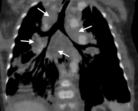
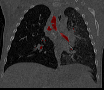
Coronal CT slice showing segmented airway regions
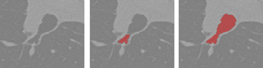
Morphological filtering a) region of an axial slice b) segmentation after coronal and sagittal filtering c) segmentation after axial filtering
Mesh representation of airway segmentation
Click and drag figure to rotate. Scroll to zoom.
Click on figure and press 'm' to toggle mesh views.
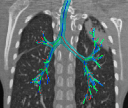
Airway segmentation and labelled skeleton. Colours are used to distinguish branches in the skeleton and a background CT slice is provided to demonstrate position
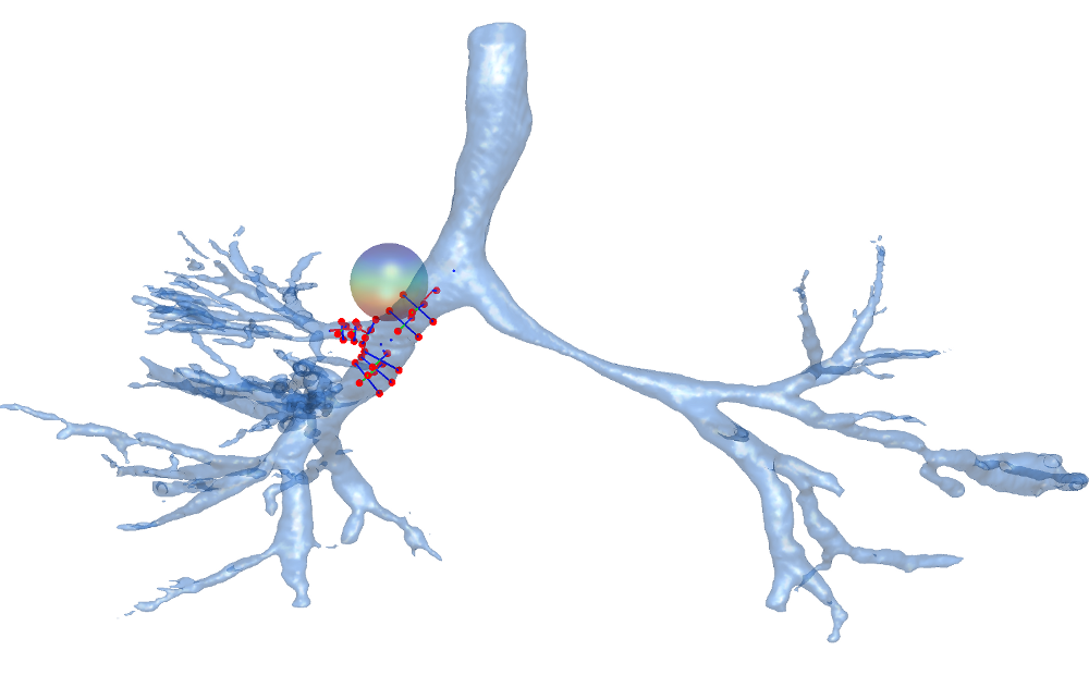
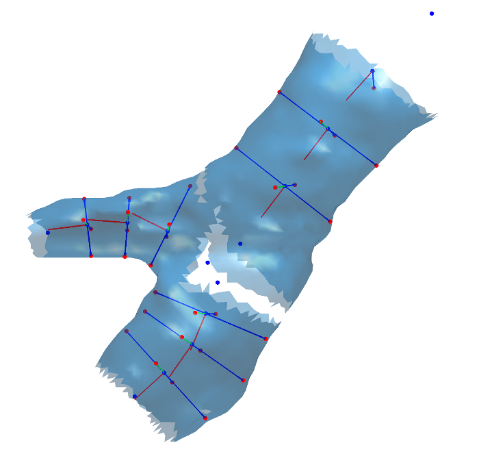
Generating surface landmarks in the region of interest on the airway surface
Registration of a template mesh to each airway region using thin-plate-spline and closest point alignment
Slider for each transform
Move each slider to the right to perform template warp
Click and drag figure to rotate. Scroll to zoom.
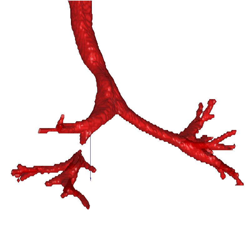
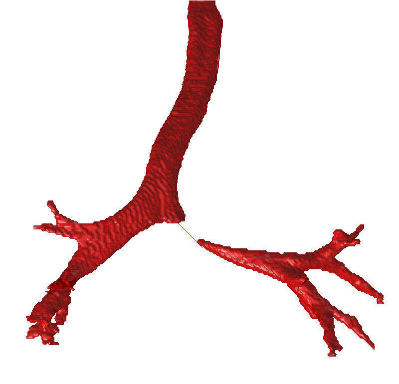
Detecting and segmenting beyond points of obstruction
A single PCA mode of variation for the airway dataset
PCA mode 1
(+ λ → 0 → - λ)
Click and drag figure to rotate. Scroll to zoom.
Move the slider to show the variation along one statistical mode
A number of modes of variation are used to train a classifier to detect airway pathology
ROC curves for classification of paediatric TB from airway shape deformation
Projection of silhouette edge vertices onto a 2D surface.
Used to assist in the segmentatio of the airways in 2D radiographs